
This comprehensive lab manual introduces students to hands-on activities and experiments in human anatomy and physiology. It includes detailed explanations, applied exercises, and essential concepts to enhance understanding of the human body’s structure and function.
Welcome to the Lab Manual for Human Anatomy and Physiology! This manual is designed to guide students through hands-on activities and experiments that enhance their understanding of the human body’s structure and function. It serves as a comprehensive resource for both classroom and laboratory settings, providing detailed explanations, practical exercises, and real-world applications of anatomical and physiological concepts.
Each section of this manual is carefully structured to align with course objectives, ensuring a seamless learning experience. From microscopic anatomy to gross dissection, and from physiological measurements to histological examinations, this manual covers a wide range of topics essential for undergraduate studies. The activities are designed to foster critical thinking, observation, and scientific inquiry, helping students develop a deeper appreciation for the complexities of the human body. Whether you’re identifying anatomical structures, conducting experiments, or analyzing data, this manual will serve as your go-to guide for mastering the principles of human anatomy and physiology.
Essential Laboratory Equipment and Tools
The anatomy and physiology lab is equipped with a variety of tools and instruments to facilitate hands-on learning. Essential equipment includes dissection kits, consisting of scalpels, forceps, and probes, which are used for examining anatomical structures. Microscopes are critical for studying histological slides, allowing students to observe tissues and cellular structures in detail. Measuring tools, such as calipers and thermometers, are used to collect physiological data, while skeletons and bone models provide a three-dimensional understanding of the skeletal system.
Other key tools include specimen trays for organizing materials, gloves and goggles for safety, and lab coats to protect clothing. First aid kits are also available to address minor accidents. Additionally, software tools, such as Anatomy and Physiology Revealed (APR), are integrated into the lab to enhance learning through digital visualization and interactive exercises. These resources ensure students are well-equipped to explore and understand the intricacies of the human body.
Safety Protocols and Precautions in the Anatomy Lab
Safety is paramount in the anatomy lab to ensure a secure and healthy learning environment. Students must adhere to specific protocols to minimize risks associated with handling biological specimens, chemicals, and sharp instruments. Essential safety measures include wearing personal protective equipment (PPE), such as lab coats, gloves, and goggles, to protect against exposure to preservatives and biological materials.
Proper handling of sharp instruments, like scalpels and needles, is critical to prevent injuries. Students should follow correct dissection techniques and ensure all instruments are stored safely after use. Additionally, chemical safety is emphasized, with proper ventilation and handling of preservatives like formaldehyde. In case of spills or exposure, students must immediately rinse affected areas and seek assistance.
Emergency preparedness includes knowing the location of fire extinguishers, eye wash stations, and first aid kits. Regular safety drills and briefings are conducted to ensure preparedness for potential incidents. By following these guidelines, students can engage in lab activities confidently while maintaining a safe environment for everyone.
Microscopic Anatomy: Techniques and Procedures
Microscopic anatomy involves the study of tissues and cells, requiring precise techniques and procedures to prepare and examine specimens. Proper slide preparation is essential, including fixation, staining, and mounting to ensure clear visibility under the microscope. Students learn to handle brightfield microscopes, focusing on proper use of objective lenses and condensers to optimize image clarity.
Key steps include placing slides on the stage, adjusting the coarse and fine focus knobs, and using the iris diaphragm to regulate light. Proper handling of slides is emphasized to prevent damage or contamination. Techniques for identifying cellular structures, such as nuclei, mitochondria, and cytoplasm, are also covered.
Advanced methods include histological staining to differentiate tissue types, such as hematoxylin and eosin (H&E) for identifying nuclei and cytoplasm. Students also learn to distinguish between normal and abnormal cellular features, enhancing their understanding of microscopic anatomy. These skills are crucial for correlating microscopic findings with gross anatomical observations.
Gross Anatomy: Dissection and Identification of Structures
Gross anatomy focuses on the study of large-scale body structures visible to the naked eye. This section provides detailed guidance on dissection techniques, emphasizing proper use of tools like scalpels, forceps, and probes. Students learn to systematically identify organs, muscles, and other anatomical features, correlating them with textbook descriptions and diagrams.
Safety protocols are highlighted, including proper handling of cadaveric material and personal protective equipment. The manual outlines a systematic approach to dissection, ensuring students understand spatial relationships between structures. Tips for distinguishing tissues and organs through color, texture, and location are also included.
Key activities include regional dissection of the torso, limbs, and head, with step-by-step instructions for exposing and identifying structures. The section emphasizes the importance of meticulous observation and accurate labeling to reinforce anatomical knowledge. By mastering these skills, students gain a robust foundation in gross anatomy, essential for advanced studies in medicine and healthcare.
Physiology Experiments: Measuring Vital Signs and Body Functions
This section of the lab manual focuses on hands-on experiments to measure and analyze vital signs and physiological processes. Students learn to accurately record heart rate, blood pressure, respiratory rate, and body temperature using tools like ECG, sphygmomanometers, and thermometers. Experiments are designed to demonstrate how these measurements reflect the body’s functional state under different conditions, such as rest or exercise.
Activities include spirometry to assess lung function, electrocardiography to study heart rhythms, and tests to evaluate reflexes and nervous system responses. The manual provides step-by-step protocols for setting up equipment, collecting data, and interpreting results. Tips for minimizing experimental errors and ensuring accurate measurements are emphasized.

These experiments help students understand the interplay between physiological systems and how external factors influence bodily functions. By conducting these exercises, learners develop practical skills in data collection and analysis, essential for careers in healthcare and biomedical sciences. The section reinforces theoretical concepts with real-world applications, making complex physiology accessible and engaging.
Histology: Examining Tissues Under the Microscope
This section of the lab manual introduces students to the study of histology, focusing on the microscopic examination of human tissues. It provides detailed protocols for preparing and staining tissue samples, ensuring clear visualization under a microscope. Essential techniques include fixation, sectioning, and staining to preserve tissue structure and enhance contrast for accurate observation.
Students learn to identify and differentiate between the four primary tissue types—epithelial, connective, muscle, and nervous—by examining their unique cellular arrangements and structures. The manual includes high-quality micrographs and diagrams to aid in recognizing key histological features. Practical exercises guide learners in using microscopy tools, such as focusing, adjusting lighting, and identifying structures at different magnifications.
Activities emphasize the importance of proper slide preparation and the role of histology in understanding disease diagnosis and tissue function. By mastering these skills, students gain a deeper appreciation for the microscopic organization of tissues and their roles in maintaining bodily functions. This section bridges theoretical knowledge with practical observation, enhancing students’ ability to analyze and interpret histological specimens.
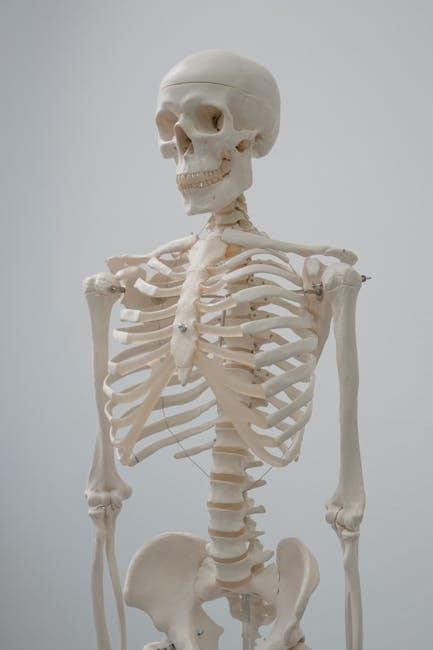
Skeletal and Muscular System: Practical Identification and Analysis
This section of the lab manual focuses on the hands-on identification and analysis of the skeletal and muscular systems. Students learn to recognize and label major bones, including their structural features and anatomical landmarks. Practical exercises involve palpation of muscles and joint movements to understand their functional roles in the body.
Key activities include identifying bone types (long, short, flat, irregular, and sesamoid) and analyzing muscle attachments and actions. Students also explore the relationship between bones and muscles, focusing on how they work together to enable movement and provide structural support. Detailed diagrams and 3D models are used to reinforce understanding of complex anatomical relationships.
Exercises such as muscle dissection and joint movement analysis help students correlate anatomical structures with physiological functions. This section emphasizes the importance of understanding the skeletal and muscular systems in maintaining posture, facilitating locomotion, and supporting daily activities. By mastering these practical skills, students gain a strong foundation for further study in anatomy and physiology.
Nervous and Circulatory Systems: Hands-On Exploration
This section of the lab manual provides hands-on activities to explore the nervous and circulatory systems. Students engage in practical exercises to identify key structures and understand their functions. For the nervous system, activities include examining histological preparations of nerve tissue and conducting reflex tests to observe neural responses. The circulatory system is studied through measurements of blood pressure, heart rate, and blood vessel structure analysis.
Students use specialized equipment, such as ECG machines, to record and interpret electrical activity of the heart. Dissection exercises reveal the organization of blood vessels and their distribution throughout the body. Interactive simulations and software tools, like Biopac Student Lab PRO, allow students to visualize and analyze physiological data, such as nerve impulses and blood flow dynamics.

Practical tasks emphasize the integration of the nervous and circulatory systems in maintaining homeostasis. By correlating anatomical structures with physiological functions, students gain a deeper understanding of how these systems interact to regulate body functions, such as blood pressure and oxygen delivery.

Lab Report Writing and Data Interpretation
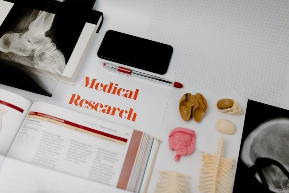
Lab report writing and data interpretation are essential skills for effectively communicating scientific findings. This section guides students in structuring clear, concise lab reports, emphasizing the importance of accurate documentation and analysis. Key components include the title, introduction, materials and methods, results, and discussion sections.
Data interpretation focuses on translating numerical and qualitative observations into meaningful conclusions. Students learn to identify patterns, calculate statistical significance, and draw logical inferences from experimental data. Graphs, charts, and tables are used to visually represent findings, enhancing clarity and understanding.
Practical exercises involve analyzing sample data sets, practicing statistical calculations, and crafting coherent interpretations. Emphasis is placed on avoiding bias and ensuring objectivity in data presentation. By mastering these skills, students develop the ability to critically evaluate experimental outcomes and articulate their significance in the context of human anatomy and physiology.